
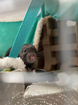
Anatomy & Physiology
From whiskers to tail, rat biology is remarkably well understood thanks to their roles in research. This page highlights the systems that keep them running.
General Appearance
Healthy rats have long, slender bodies with mostly hairless tails. These tails can be as long as 85% of the total body length, with females having longer tails proportional to their body than males. Although the tail appears hairless, it is covered in small, overlapping scales and fine hairs. These scales aid in allowing rats to grip surfaces when wrapping the tail around nearby objects to balance or support themselves. The overlapping structure ensures the tail can easily bend and flex without causing damage to the skin underneath.

Rat tail scales
A rat’s body is covered with fur except on the nose, lips, feet, and tail. Rats have several types of hair that serve different purposes, including protection, sensing touch, and insulation. Skin is loose and flexible, helping with movement and body function.
Both the front and hind paws have five digits. The first digit on the front paw is smaller and resembles a nub.

Note the tiny nub on the front paw
Sexual Dimorphism
Male and female rats differ in both size and reproductive anatomy. Standard males usually weigh between 400 and 700g, whereas females are on the smaller end sitting between 200 and 400g. However, size is heavily dependent on genetic lines.
Females have 12 nipples arranged in six pairs along the underside of the body. Male rats do not have nipples but they still possess mammary tissue.

Male Rat
Credit: @jasminep04

Female Rat
Credit: @antares_anta
In females, the vaginal opening is located just below the anus, with the clitoris sitting at the front of the opening. In males, the scrotum is located toward the rear underside of the body. This may change with appearance though, as they can retract testes when stressed. The penis lies within a sheath called the prepuce and contains a small bone known as the os penis.

Urogenital Distance Comparisons between males and females
Credit: Sweet Whispers Rattery
Musculoskeletal System
The rat musculoskeletal system is similar to that of other four-legged mammals and is divided into the axial skeleton (skull, spine, and ribs) and the appendicular skeleton (limbs and limb girdles). The vertebral column consists of 7 cervical, 13 thoracic, 6 lumbar, 4 sacral, and 27–30 caudal (tail) vertebrae. Bone growth is most rapid around 7 weeks of age and generally ceases by about 6 months. Skeletal muscle is formed from bundles of muscle fibres connected by connective tissue and attached to bone via tendons. Within each fibre, myofibrils generate contraction and enable movement. Muscle strength and endurance decline with age, as in other mammals.
Systemic regulation of skeletal growth and metabolism is strongly influenced by the endocrine system, including the pituitary gland. Located at the base of the brain, the pituitary secretes key hormones such as growth hormone, which regulates bone growth, protein synthesis, and overall body development. It also interacts with other endocrine organs, including the thyroid and adrenal glands, making it a central regulator of growth and physiological balance.
Pituitary tumours are a relatively common finding in rats, particularly in older animals, with incidence varying widely depending on strain, sex, and age. These tumours typically arise from hormone-producing pituitary cells, but they may grow and compress surrounding brain structures or disrupt endocrine signalling. As a result, affected animals may show either neurological signs from local pressure effects or hormonal disturbances due to disruption of the hypothalamic–pituitary axis.
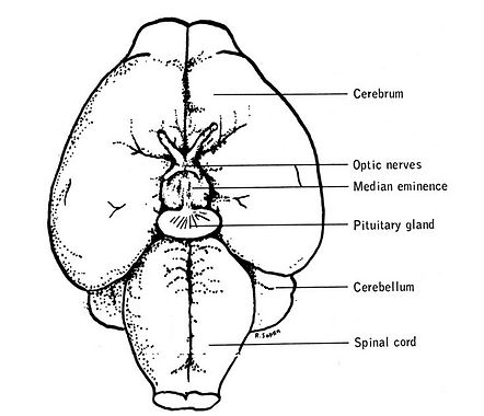
The Rat Brain
Credit: Jax Laboratories
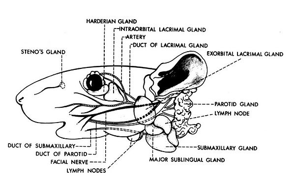
In the head and neck region, several glands support key physiological functions. Rats have two lacrimal glands and a Harderian gland, which help lubricate and protect the eye. The Harderian gland produces tears rich in porphyrin pigments, giving tears a reddish colour. Excess porphyrin around the eyes or nose can be a sign of stress, illness, dehydration, or poor welfare. Nasal glands help humidify inhaled air and contribute to respiratory defence. Immune organs such as lymph nodes and the thymus are also present in this region.
Superficial glands of the head and neck (lateral aspect). Redrawn from Greene, E.C., 1935. Anatomy of the Rat, vol. 27. American Philosophical Society, Philadelphia, PA.
Rats possess both white and brown adipose tissue. White fat primarily stores and supplies energy, whereas brown fat generates heat through thermogenesis and plays an important role in energy balance, metabolism, and obesity research.
Sensory Systems
Vision
The eyes of rats are similar to other mammals and consist of an iris, cornea, lens, and retina. The cornea is a clear outer layer that helps focus light and has a stronger refractive power than the lens itself. The iris controls how much light enters the eye by changing the size of the pupil.
In albino rats, the iris lacks pigment and appears almost transparent, making the retina much more sensitive to bright light. Because of this, albino rats are particularly prone to light-induced retinal damage. Rats generally prefer dim environments, and prolonged exposure to bright light can damage the retina. Low to moderate light levels are recommended, and it is advised to never take photos or videos of rats with the flash on.
Unlike humans, rats have poorly developed ciliary muscles, meaning they have very limited ability to adjust focus. Pigmented rats have vision roughly equivalent to 20/600, while albinos have even poorer vision at around 20/1200. Despite this, rats have a wide field of view that helps them detect movement and predators. Their vision is adapted more for low-light and nocturnal activity than for detailed image resolution.
Although they were once thought to be colour blind, they actually possess two types of cone cells and are capable of limited colour vision, especially in the blue and green ranges of the spectrum.
Smell
Rats rely heavily on scent for communication and survival. Odours help them identify other rats, recognise family members, even determine social rank. Air entering and leaving the nose is directed sideways through each nostril, reducing mixing of scents and allowing rats to sample odours separately from each side of the nose.
Inside the nasal cavity are several types of specialized epithelial tissues. About half of the nasal surface is covered by olfactory epithelium, which contains millions of olfactory sensory neurons used to detect odours. The nose also contains mucus-producing goblet cells and Bowman’s glands, which help dissolve odour molecules and protect the nasal tissues.
Taste
Just like humans, rats can detect taste using taste buds located on the tongue, hard and soft palate, and pharynx. Taste buds are small oval-shaped sensory organs made up of supporting tissue and specialized receptor cells known as gustatory cells. These receptor cells have tiny hair-like projections that extend into an opening called the taste pore, where they come into contact with dissolved food particles.
Taste buds contain several different cell types with specific functions. Type I cells mainly act as supporting cells and may help detect salty tastes. Type II cells detect sweet, bitter, and umami flavours, while Type III cells are involved in sensing sour tastes as well as some sweet, bitter, and umami signals.
Hearing
Rats have very sensitive hearing and can detect ultrasonic sounds up to around 80,000 Hz, far beyond the range of human hearing. These ultrasonic vocalizations are important for communication, especially during social interactions. The ear consists of the external, middle, and inner ear.
The external ear collects sound and directs it toward the tympanic membrane (eardrum). At the base of the ear is the Zymbal’s gland, a modified sebaceous gland that secretes into the ear canal. This gland is important clinically as it is a common site of tumour formation in rats.
The middle ear contains the auditory ear bones (ossicles) which transfer sound vibrations to the cochlea in the inner ear. Inside the cochlea, specialized hair cells convert sound waves into nerve signals that travel to the brain. The inner ear also contains the vestibular system, including the vestibule and semicircular canals, which control balance and spatial orientation by detecting movement and changes in head position.
Touch
Rats rely heavily on their whiskers (vibrissae) for touch and environmental awareness. The most noticeable whiskers are the mystacial vibrissae on the snout, although additional whiskers are located above the eyes, along the face and neck, under the chin, and on the forelimbs. Each whisker sits within a specialized follicle surrounded by a blood-filled sinus and sensory nerves.
When a whisker touches an object, it bends and stimulates nerve endings that send signals to the brain. Rats use their whiskers to navigate, detect objects, maintain balance, and interact socially, especially in low-light environments. The whiskers move rhythmically under muscular control, allowing rats to actively explore their surroundings through touch. The nose and whiskers together form the primary system for tactile and sensory exploration in rats.
Dentition and Jaw Structure
Rats have a highly specialised gnawing dentition adapted for continuous growth and high wear. The dental formula is 2(I 1/1, C 0/0, PM 0/0, M 3/3) = 16. The two pairs of front teeth (incisors) are open-rooted and constantly growing throughout their lifetime. The three pairs of back teeth (molars) remain closed-rooted and do not. Unlike humans, a rodent's lower jaw (mandible) is divided into two parts, the left and right mandibular bones, and can separate from one another when grinding down incisors.

Rat Skull and accompanying jaw bones
Digestive System
Within a 24-hour period, a standard rat consumes approximately 5g of food and drinks 10mL per 100g of body weight. They are omnivores, consuming a high fibre, low-fat diet primarily consisting of grains.
Following initial digestion of food in the stomach, bile assists in the breakdown of fats and elimination of waste products . Rats do not have a gallbladder, so this is produced continuously by the liver and pancreas. Further along the tract, the caecum is home to large numbers of beneficial bacteria that ferment plant material, producing additional nutrients and aiding digestion.
A key feature of rats is that they are incapable of vomiting, due to anatomical and neurological constraints.

The Digestive Tract of the Rat
Credit: Usyal, M. (201). Caecum location in laboratory rats and mice: an anatomical and radiological study. Sage Journals.
Respiratory System
Rats are obligate nasal breathers due to the close anatomical relationship between the soft palate and epiglottis, which prevents open-mouth respiration during normal breathing. A healthy rat will never open-mouth breathe.
The upper respiratory system of the rat is very similar to that of other mammals. Air enters through the external nares and passes into the nasal passages, then into the nasopharynx, where structures such as the Eustachian tubes connect the throat region to the middle ear. The airway continues past the epiglottis (which helps protect the entrance to the trachea during swallowing) into the trachea. The inner lining of the airways is specialised for protection and clearance, moving mucous and trapped particles upward towards the mouth through the cilia and producing mucous and antimicrobial secretions. This “mucociliary escalator” forms the most important first-line defense against dust, pathogens, and irritants.
In the lower respiratory system, the trachea divides into two main bronchi that supply the lungs. Rat lungs are divided into five lobes (one on the left and four on the right). These bronchi branch into progressively smaller airways, ending in microscopic air sacs where oxygen and carbon dioxide exchange occurs (alveoli).

Lung Anatomy of the Rat
Credit: Nawata et al., (2001). Sequential Bilateral Isolated Lung Perfusion in the Rat: An Experimental Model. The Annals of Thoracic Surgery.